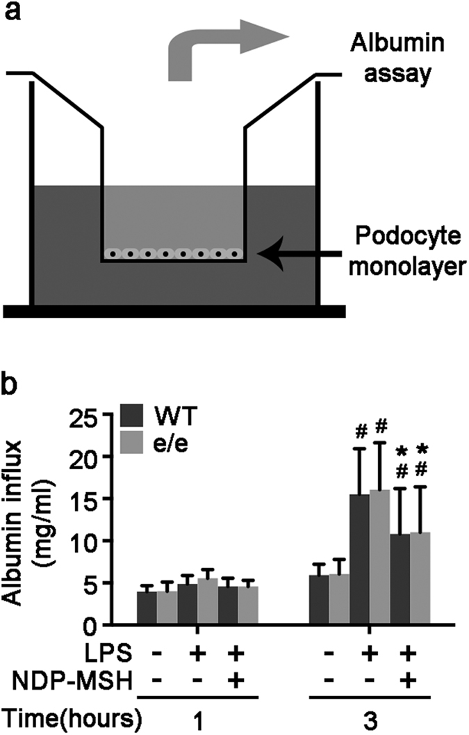
Figure 6. MC1R is not required for the protective effect of NDP-MSH on the LPS-impaired filtration barrier function of podocyte monolayers.

Primary podocytes were prepared from glomeruli isolated from MC1Re/e mice and wild-type (WT) littermates. Following different treatments, paracellular permeability assay was carried out to determine the filtration barrier function of podocytes monolayers. (a) A simplified schematic representation of the paracellular permeability assay adopted to assess the filtration barrier function of podocytes monolayers. Podocyte monolayers on collagen-coated Transwell filters were injured with LPS (20 μg/ml) or saline in the presence or absence of NDP-MSH (10−7 M) for 3 hours, and albumin permeability across podocyte monolayers was then determined. (b) Quantification of the albumin influx across podocyte monolayers. Duration of albumin incubation is shown on x-axis. #P < 0.01 vs control group (n = 6); *P < 0.05 vs LPS group (n = 6).
