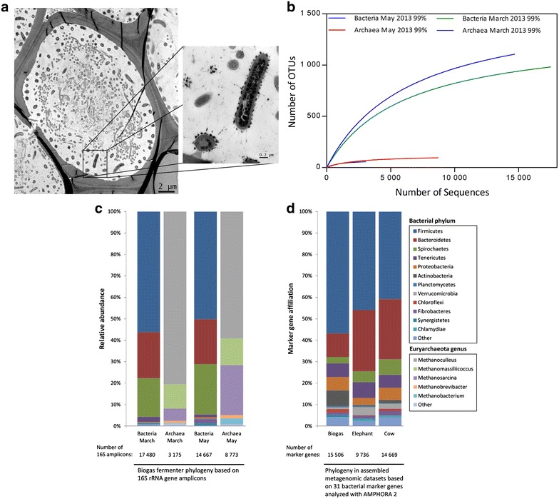
Fig. 1.

a TEM micrograph of a decomposing plant cell and the associated microorganisms in a biogas fermenter. Cellulosome-producing bacteria are almost exclusively observed in close association with the plant cell wall, where they appear to suppress growth of other microbes. Most other microorganisms are located at the more central part of the decomposing plant cell. Cellulosome-producing bacteria were identified by the large dark spots attached to the cells. b Rarefaction curves calculated for two fermenter samples of the studied agricultural biogas plant. The OTUs were clustered at 99 % genetic similarity of 16S rRNA genes. The sequences were denoised employing Acacia, and chimeric sequences were removed using UCHIME. Singleton OTUs were removed prior to the rarefaction analysis. c Phylogenetic analysis of two biogas fermenter samples based on 16S rRNA gene amplicons. The bars indicate the relative abundance of bacterial phyla and euryarchaeota genera in two samples taken from the same fermenter at different time points (March and May 2013). d Phylogenetic analysis of three assembled metagenomic data sets based on 31 bacterial marker genes. The bars show the marker gene affiliation to bacterial phyla in the data sets derived from biogas fermenter May sample and for reasons of comparison from published data sets of elephant feces and cow rumen samples [15, 23]. For this analysis, the AMPHORA 2 software was used
