
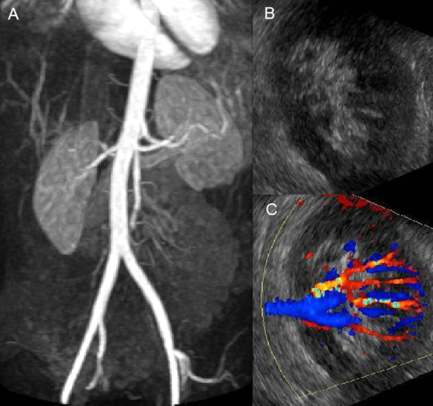
Figure 2.
27-year-old woman with acute renal infarction. (A) Coronal MRI shows perfusion defects in the lower pole of the left kidney. (B) Sonogram shows normal left kidney. (C) Doppler shows no renovascular abnormality.
Official websites use .gov
A
.gov website belongs to an official
government organization in the United States.
Secure .gov websites use HTTPS
A lock (
) or https:// means you've safely
connected to the .gov website. Share sensitive
information only on official, secure websites.
27-year-old woman with acute renal infarction. (A) Coronal MRI shows perfusion defects in the lower pole of the left kidney. (B) Sonogram shows normal left kidney. (C) Doppler shows no renovascular abnormality.