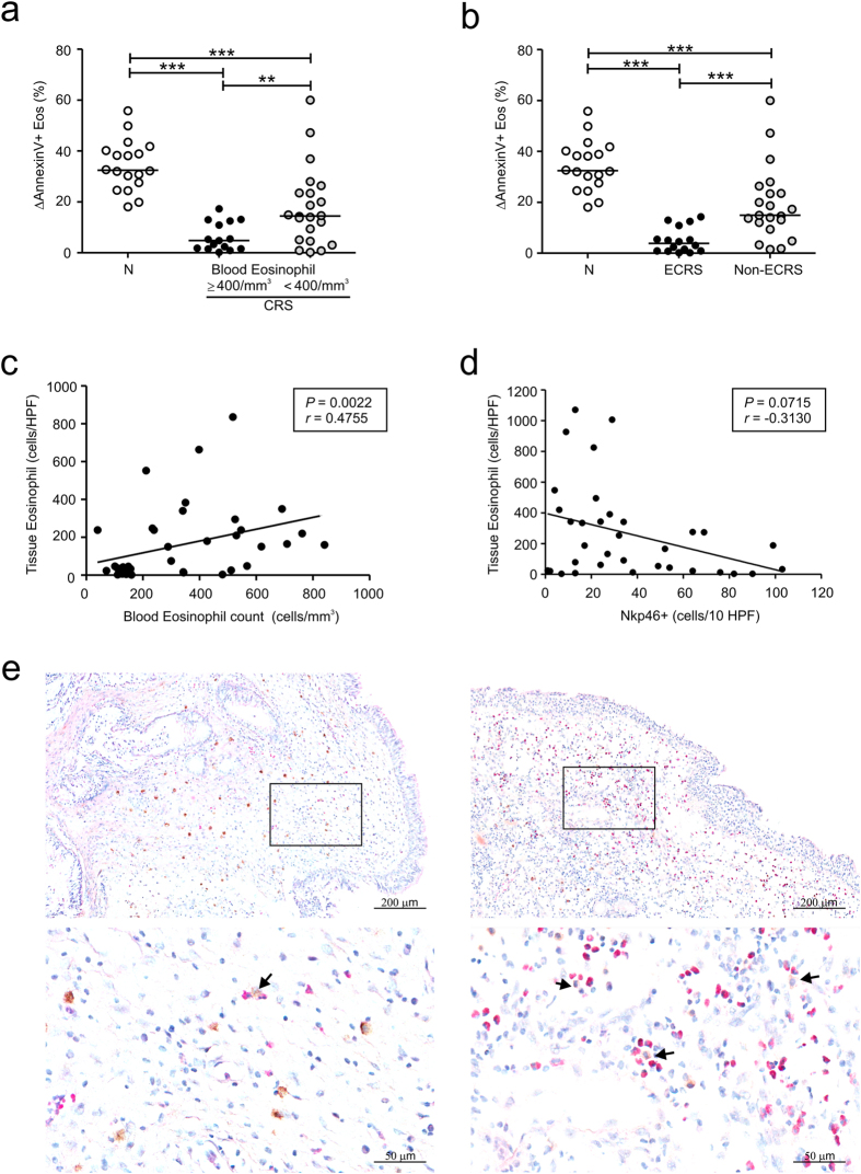
Figure 2. Eosinophilic inflammation is associated with reduced NK cell function in CRS.
(a,b) Patients with CRS were divided into eosinophilic (n = 15) and noneosinophilic (n = 22) groups on the basis of blood eosinophil counts (a) or eosinophilic CRS (ECRS) (n = 16) and non-ECRS (n = 21) groups based on tissue eosinophil counts (b). PBMCs from each group were incubated with autologous peripheral blood granulocytes. The three groups were compared via statistical dot plots in terms of the relative percent increase in eosinophil apoptosis (ΔAnnexin V+ Eos). (c) The peripheral blood eosinophil counts correlated positively with the frequency of eosinophil counts in sinus tissue from CRS patients (n = 39). (d) The counts of NKp46+ NK cells correlated inversely with the frequency of eosinophil counts in sinus tissue from CRS patients (n = 34). (e) Representative immunostaining for NKp46+ NK cells and hematoxylin and eosin counterstaining in sinonasal tissue from patients with non-ECRS (left panel) and ECRS (right panel). Arrows denote NKp46+ NK cells in contact with or close to eosinophils. Scale bars represent 200 μm (top panel) and 50 μm (bottom panel). Horizontal bars denote the medians. ***P < 0.001, Mann-Whitney U test (a,b) and Spearman correlation test (c,d).

