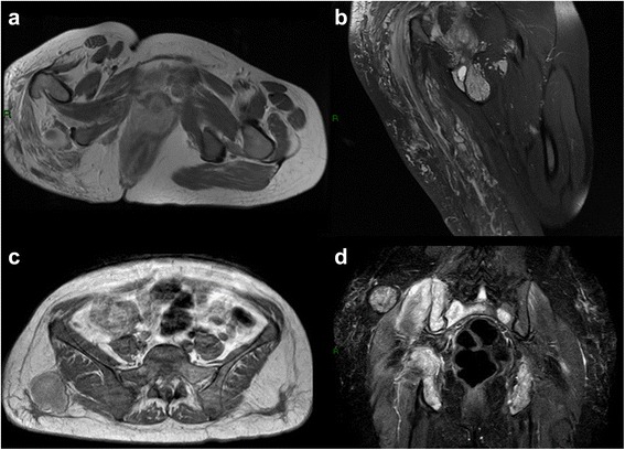
Fig. 4.

Traditional MR imaging. Axial T1 with contrast media and coronal DP fat of huge angiomatosis of the gluteal region and upper right tight. Either in T1 with contrast or in DP fat weighted image is possible to recognize the huge, mingled, and interspersed, vessels proliferation (a-b). Metastasis of angiosarcoma of the breast involving either the soft tissue of the gluteal region or the bone of sacral and iliac wing (c-d), axial T1 with contrast media and coronal STIR. In T1 only the soft tissue lesion is hyperintense, in STIR both lesion, soft tissue and bone are hyperintense
