Figure 4.
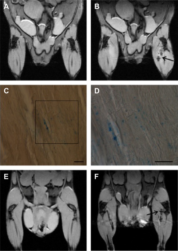
In vivo MRI of exosomes-USPIO.
Notes: In vivo MR images acquired preintramuscular (A) and postintramuscular (B) injections of exosomes-USPIO (arrow). Prussian blue histological examination of extracted muscle tissue: blue spots inside the muscle confirmed the presence of iron nanoparticles (C) (magnification ×20, scale bar 50 μm). D shows a higher magnification (×40, scale bar 50 μm) of the boxed area shown in C. In vivo MR images acquired preintramuscular (E) and postintramuscular (F) injections of plain USPIO (arrow) containing the same amount of iron of labeled exosomes; the signal is comparable with that detected in exosomes-USPIO (B).
Abbreviations: MR, magnetic resonance; MRI, magnetic resonance imaging; USPIO, ultrasmall superparamagnetic iron oxide nanoparticles.

