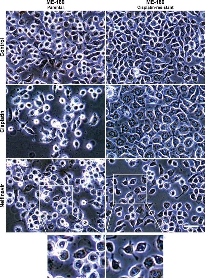
Figure 1.
Morphology of ME-180 cells with or without nelfinavir treatment.
Notes: Cells were treated with control 0.1% dimethyl sulfoxide or with 10 μM nelfinavir after 20 hours. While some proliferation is inhibited by nelfinavir by this time point, as indicated by lower cell confluence, the cellular morphology is relatively unchanged. ME-180 cells often have an obvious vacuole, which tends to increase in size following treatment with nelfinavir. The scale bar indicates 30 μm and applies to all images with the exception of the two magnified excerpts shown at the bottom of the figure.

