Figure 2.
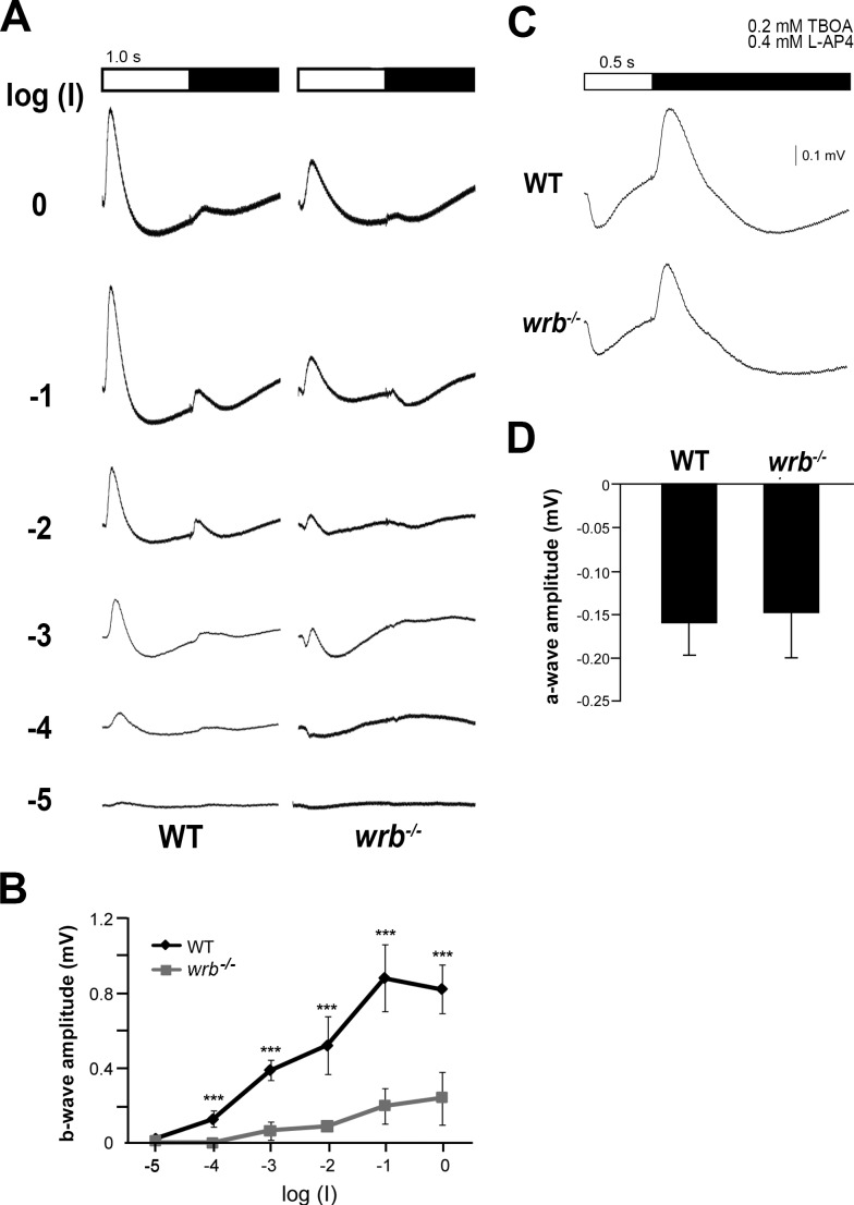
ERG reveals disrupted outer retina signaling in wrb−/− mutants. (A) Averaged ERG traces from wild-type and wrb−/− larval eyes elicited by a series of white flashes with onset and duration depicted at top. Flash intensity was incremented by log unit steps from bottom to top with log(I) = −1 corresponding to 5.3 × 10³ μW/cm2 at 500 nm. The interstimulus interval was 10 seconds. (B) Response versus stimulus functions for average peak b-wave amplitudes from wild-type and wrb−/− larvae, as measured from a-wave trough to b-wave peak. (C) Individual ERG traces elicited by flashes after treatment with TBOA and L-AP4 to eliminate ERG components arising from glutamate-dependent signaling. Each flash was 0.5 seconds in duration and corresponded to log(I) = −1 intensity. (D) Average a-wave maximum amplitudes from wild-type and wrb−/− larvae (n = 10, wild-type, n = 10, wrb−/−). Error bars denote SEM. Significance levels are as follows: ***P < 0.0001.

