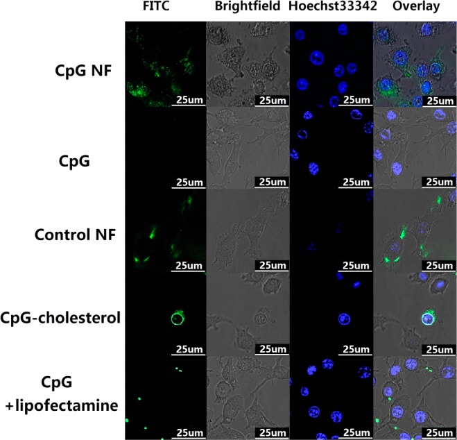
Figure 3.

Confocal images showing the internalization of NFs by RAW264.7 macrophages. Confocal microscopy images showing that CpG NFs (100 nM CpG equivalents), as well as control NFs, were internalized into RAW 264.7 macrophages after incubation for 2 h. NFs were incorporated with FITC (green) by attaching it to the 5′ end of the RCR primer. 3′-end-FITC-labeled CpG ODNs were used as a negative control group. Internalization of FITC–CpG–cholesterol conjugation and lipofectamine-mediated CpG FITC was also investigated and compared.
