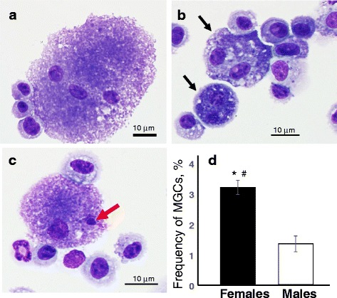
Fig. 4.

Representative light micrographs of giant multi-nucleated (a-c) cells including bi-nucleated (b) and micro-nucleated (c) alveolar microphages from BAL fluids of female mice 3 month post repeated exposure with CNC (marked by arrows). Frequency of giant multi-nucleated cells (MGCs) in BAL fluids of female or male mice 3 month after the last exposure with CNC (d). Black columns – giant BAL cells from female mice exposed to CNC, open columns– giant BAL cells from male mice exposed to CNC. Mean ± SEM (n = 10 mice/group). *p < 0.05, vs control PBS-exposed mice, # p < 0.05, vs male mice exposed to CNC. Blind-coded slides were independently scored by two readers. A total of 2000 cells per sample were scored. No MGCs were found in control mice
