
Fig. 5.
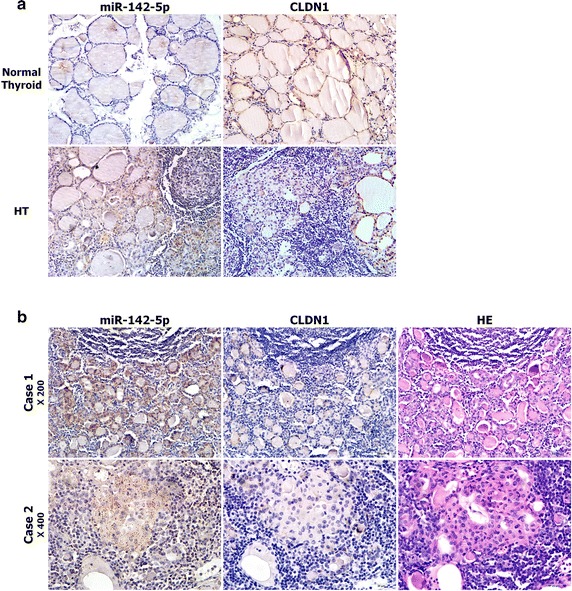
Immunostaining of claudin-1 (CLDN1) compared with the ISH of miR-142-5p in FFPE samples. a The mutually exclusive patterns between miR-142-5p (left) and CLDN1 (right) expression in normal thyroid and HT tissues (×200). b Images from serial FFPE HT tissue sections in which LNA-ISH for miR-142-5p (left), immunostaining for CLDN1 (middle) and HE staining (right) were done (top ×200, below ×400)