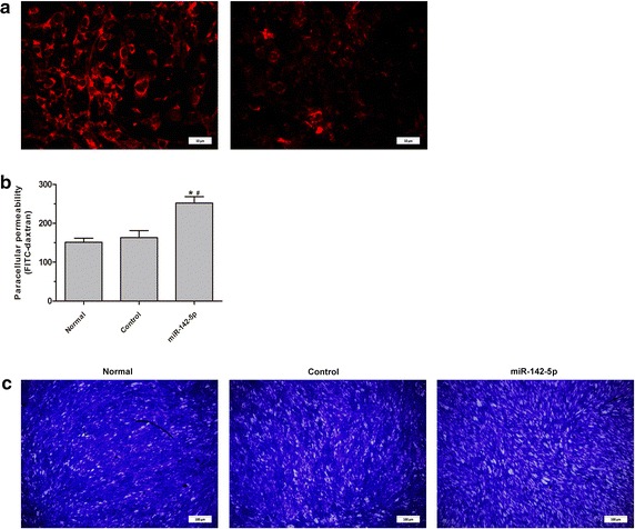
Fig. 6.

Immunofluoresence staining and permeability test of thyrocytes. a Immunofluoresence staining of CLDN1 in thyrocytes transfected with negative control lentivirus vector (linear distribution of CLDN1, left ×1000) and mir-142 lentivirus vector (discontinuous and diminished staining pattern, right, ×1000). b Permeability test of the thyrocyte monolayer, which showed the increased permeability of the monolayer thyrocytes (*P < 0.01 vs. normal, # P < 0.01 vs. control). c Cells staining of the thyrocyte monolayer after permeability testing, which showed the increased of the intercellular gap (×100)
