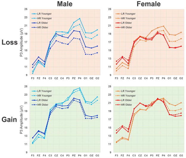
Fig. 3.
Qualitative comparison of mean P3 values for each subgroup during loss and gain conditions across 12 electrodes representing 4 regions (frontal: F3, FZ, F4; central: C3, CZ, C4; parietal: P3, PZ, P4; occipital: O1, OZ, O2). HR offspring display markedly lower P3 amplitude than LR subjects in both younger and older male subgroups during loss condition as well as in younger female subjects during both loss and gain conditions.

