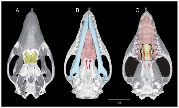
Figure 4.
Mature skull of Monodelphis reconstructed from CT data. A) dorsal view cut-away to show cribriform plate (yellow); (B) ventral view, with jaws (blue) and secondary palate (red) with arrows showing retronasal entrance to the nose via the choanae; (C) jaws and part of secondary palate removed with arrows showing the sphenethmoidal apertures in the ossified floor of the nasal capsule (yellow), which direct retronasal airflow across olfactory epithelium.

