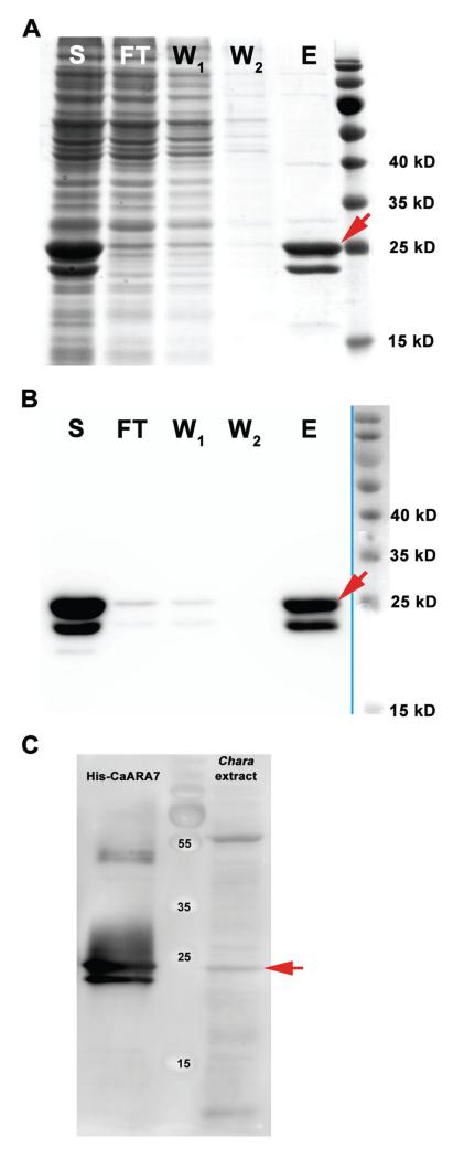
Figure 2. Purification of His-tagged CaARA7 and detection of CaARA7 in Chara protein extracts.
SDS-PAGE (A) and western blot (B) of different purification steps of recombinantly expressed Chara australis ARA7. Purification was performed by Ni-TED affinity column. Lanes are indicated as follows: S = soluble protein fraction of E. coli lysate; FT = flow through of affinity column; W1 and W2 = wash step one and two; E = eluted purified His-tagged CaARA7 protein (red arrows). Important molecular mass marker bands are indicated (right lane). C) Western blot of C. australis protein extract and purified His-tagged CaARA7. Recombinant His-CaARA7 was used for control. A polyconal antibody against ARA7 detected a prominent band with a molecular mass of about 25 kD in the protein extract of Chara (red arrow) and two bands in recombinant His-CaARA7. Important molecular mass marker bands are indicated (middle lane).

