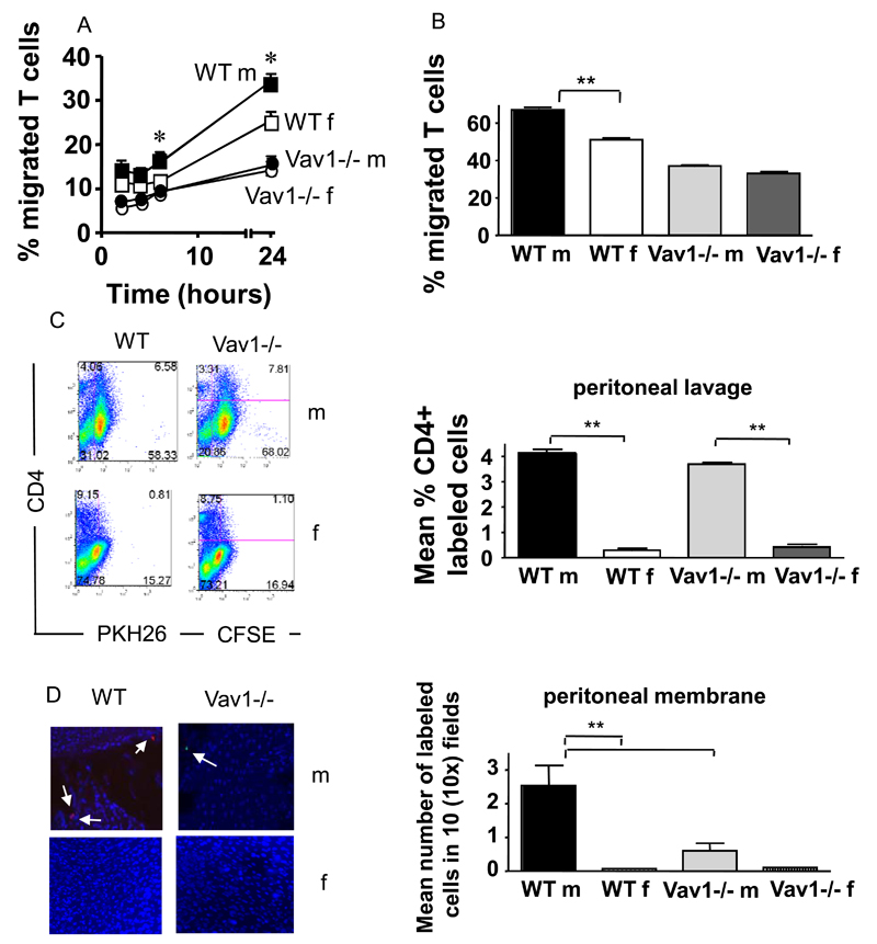
Figure 3. Antigen-driven Vav1-/- T-cell migration.
HY-specific CD4+ WT and Vav1-/- T cells (3x10⁵) were seeded onto IFNγ-treated antigenic (male) or non-antigenic (female) syngeneic EC monolayers grown on transwells. The mean percentage migration at the indicated time points from three experiments of similar design is shown. Panel B: T cells (1x10⁶) were plated on 35mm dishes coated with of IFNγ-treated antigenic (male) or non-antigenic (female) EC and allowed to migrate for 50 minutes. Pictures were taken every 30 seconds and analysed as described in Materials and Methods. Transmigrating T cells were defined as changing phase i.e. turning from bright to dark once under the endothelial monolayer. Mean percentage of cells transmigrating was calculated from three independent experiments from a sample of 100 cells per movie.
Panels C-F: HY-specific CD4+ WT and Vav1-/- T cells labeled with PKH26 (red) and CFSE (green), respectively were co-injected iv into syngeneic male or female recipients which had previously received an ip injection of IFNγ. Labeled T-cell enrichment into the peritoneal lavage was analysed by flow cytometry 24 hours later (C). The mean percentage of CD4+ labeled (PKH26 or CFSE) T cells in the peritoneal lavage from at least three animals is shown (D). Nuclei are stained by DAPI (blue). Retention of T cells into the peritoneal membrane (panel E) was analysed by wide-field fluorescence microscopy as described in the legend to Figure 2. The mean number of cells in six tissue samples from at least three mice is shown (panel F).
Error bars indicate standard error of the mean (*p<0.05, **p<0.01).

