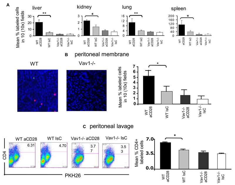
Figure 7. Vav1-/- - cell motility is not susceptible to CD28-mediated regulation.
Panel A: HY-specific CD4WT and Vav1-/- T cells which had either undergone antibody-mediated CD28 ligation (30 minutes at 37°C, PKH26-labeled) or had been pre-treated with an antibody isotype control (CFSE-labeled) were injected iv (107/mouse) into syngeneic female recipients. The presence of fluorescently labeled cells in the indicated organs was assessed 24 hours later as described in the legend to Figure 2. The mean T-cell number ± SEM observed in samples from at least six animals are shown (*p<0.05, **p<0.01).
Panel B-C: HY-specific CD4+ WT and Vav1-/- T cells, which had either undergone antibody-mediated CD28 ligation (PKH26-labeled) or had been pre-treated with an antibody isotype control (CFSE-labeled) were injected iv (107/mouse) into male mice that had received an ip injection of IFNγ 48 hours earlier. The presence of fluorescently labeled cells in the peritoneal membrane (B) and cavity (C)) was assessed 24 hours later as described in the legend to Figure 3. The mean T-cell number ± SEM observed in samples from at least three animals is shown in the right-hand side panels (*p<0.05, **p<0.01).

