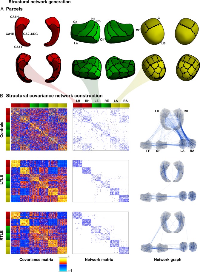
Figure 1.
Mesiotemporal circuit representation based on subregional volume covariance. (A) Low-resolution (upper panel) and high-resolution (lower panel) parcellation of the hippocampus (red), entorhinal cortex (green), and amygdala (yellow). (B) Covariance network in controls, and patients with left and right temporal lobe epilepsy (LTLE, RTLE). The left column displays high-resolution structural covariance matrices, the middle column displays the binary network matrices thresholded at a density of 8% (threshold ensuring fully connected networks in all 3 groups), the right column illustrates the corresponding network graphs. Color bars adjacent to the matrices signify the respective parcels as indicated in (A), with red colors representing the left/right hippocampus (LH/RH), green the left/right entorhinal cortex (LE/RE), and yellow the left and right amygdala (LA/RA). Hippocampus: CA, cornu ammonis; DG, dentate gyrus; H, hippocampal head; B, body; T, tail. Entorhinal cortex: Cd, caudal; Int, intermediate; La, lateral; Olf, olfactory; Ro, rostral. Amygdala nuclear groups: C, central; LB, laterobasal; MC, medial and cortical.

