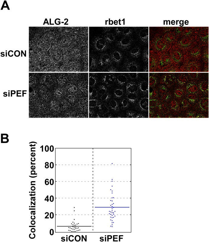
Fig 3. Peflin depletion causes increased targeting of ALG-2 to COPII membranes.
(A) NRK cells transfected with the indicated siRNA were fluorescently labeled with anti-ALG-2 antibody and cy3, or anti-rbet1 antibody (ERES and early VTC marker) and cy5. Maximum intensity projections from deconvolved widefield z-stacks are displayed in grayscale along with the corresponding pseudocolor merged panel. Increased focal and decreased diffuse labeling of ALG-2 and an increased quantity of yellow in the merged images can be observed in siPeflin cells. (B) Quantitation of multiple images. Colocalization was defined as the proportion of the total ALG-2 labeling that was coincident with rbet1 labeling, calculated for each cell individually in randomly selected fields. A T-test was performed between the siControl group and siPeflin: n = 69, p<0.0001.

