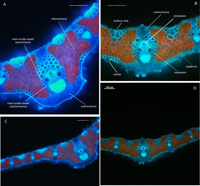
Fig 1. Brachypodium pinnatum leaf anatomy from fluorescence microscopy.
Transverse cross section of leaf blade of individuals from old (A, C) and young (B, D) populations. Notice the differences in number and size of bulliform cells occurred on the adaxial side of leaf blade. Transverse section in the midrib at median level (A, B). The differences in (i) thickness and shape of the leaf blades in zone of the central rib, width of the central rib of tiller leaf, (ii) surface, height and width of the central vascular bundle and(iii) number of the sclerenchyma strands on abaxial side of leaf and (iv) distribution of are visible. Detailed leaf measurements were presented in the Results. A, B—20× magnification, C, D—10× magnification. Bars on each of the pictures indicate 100 μm.

