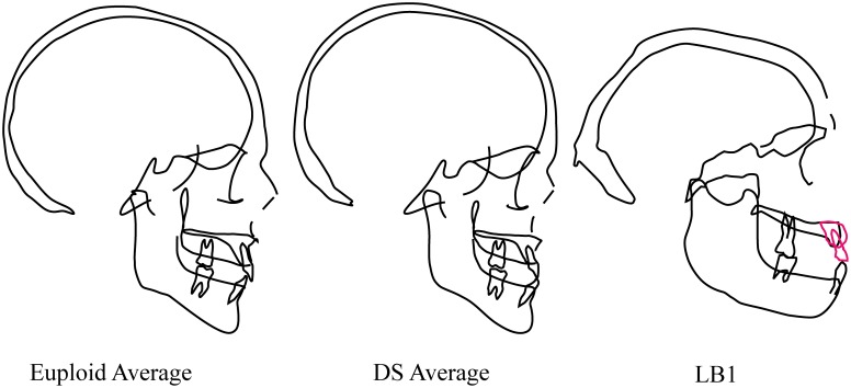
Fig 4. “Cranial templates” for euploid and DS samples of males between the ages of 19 and 29 years and pseudo-lateral cephalogram tracing of LB1.
The euploid and DS cephalograms were based on average roentgencephalometric dimensions (modified from Kisling [49]). All three images have been scaled to approximately the same cranial length. Midfacial hypoplasia in the DS facial phenotype is apparent and contrasts strongly with the relatively long and prognathic maxilla and mandible of LB1. Other differences include the thicker cranial bones, shape of the mandible and the low neurocranial profile of LB1. Note that the LB1 cranium suffered damage to midline structures of the face, including the glabella, nasal bones and subnasal region; morphology of anterior maxilla was estimated based on surrounding morphology and indication of edge-to-edge occlusion of incisors by PB.

