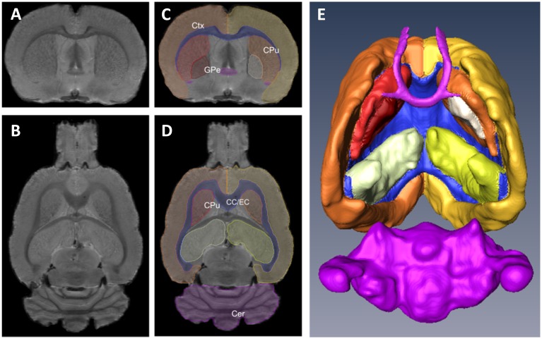
Fig 2. Rat brain MRI slices and three-dimensional reconstruction.
A-B Coronal (A) and horizontal (B) brain MRI slices from a rat brain. C-D Outlined regions of interest from the same slices. E Three-dimensional reconstruction of the segmented regions of interest; ventral view. Ctx: cerebral cortex, CPu: caudate-putamen (striatum), GPe: globus pallidus, CC/EC: corpus callosum/external capsule, Cer: cerebellum.

