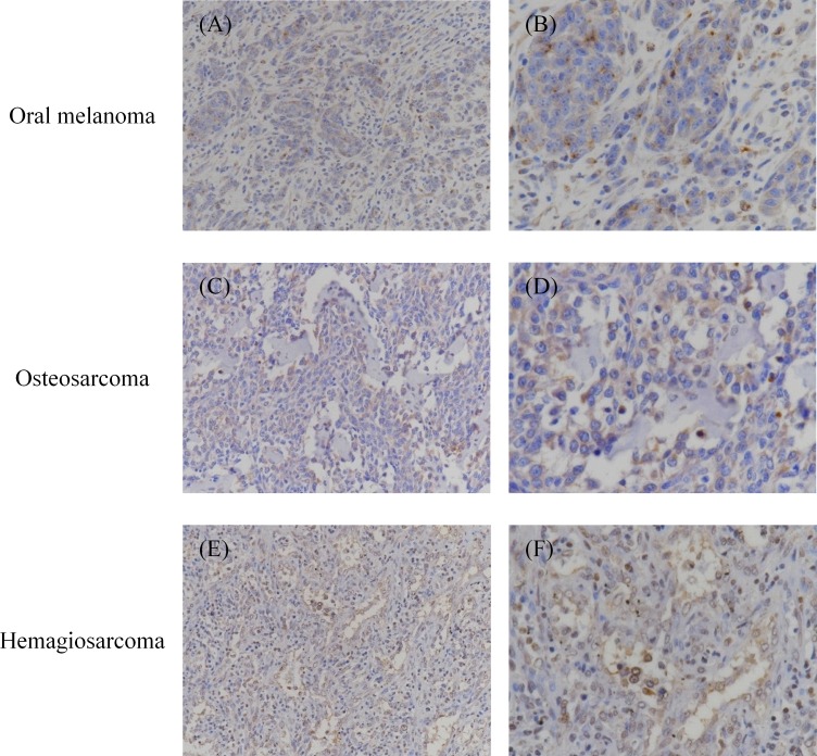
Fig 2. Immunohistochemical analysis of PD-L1 in oral melanoma, osteosarcoma, and hemangiosarcoma.
Formalin-fixed and paraffin-embedded tumor tissues were examined immunohistochemically. The sections were stained with anti-PD-L1 mAb 6G7-E1. Representative positive stainings of (A, B) oral melanoma, (C, D) osteosarcoma, and (E, F) hemangiosarcoma are shown. Original magnification: (A, C, E) 200×; (B, D, F) 400×.

