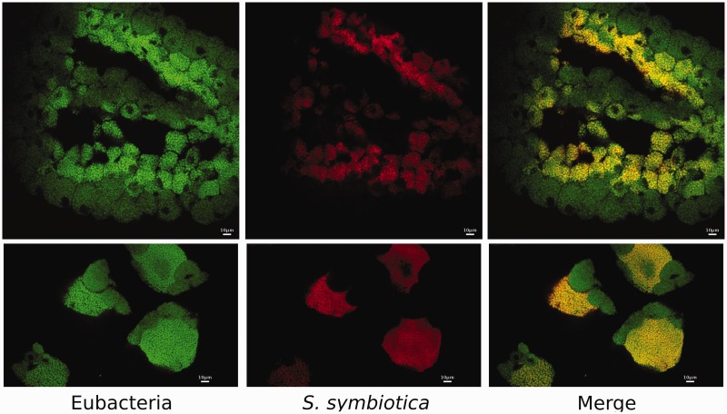
Fig. 2.—
Serratia symbiotica localization in Tuberolachnus salignus bacteriomes of early embryos. Whole-mount fluorescence in situ hybridization of early T. salignus embryos using 16S rRNA-directed probes. (Left) Eubacterial staining using EUB338 (Amann et al. 1990) 6-FAM labeled probe. (Center) S. symbiotica staining using STs (see Materials and Methods) DY-405 labeled probe. (Right) Merged image of both eubacterial and S. symbiotica staining, showing double-labeling of S. symbiotica and single-labeling of Buchnera with eubacterial probe in bacteriocytes of T. salignus in (Bottom) early and (Top) later embryos.

