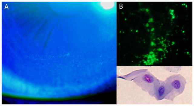
Figure 1.
(A) Photograph of the fluorescein stained right eye shows punctate staining pattern inferiorly on the cornea of a dry eye patient. (B) Fluorescent microscopy of polycarbonate filter shows singly stained cells throughout the filter (x40). (C) Polycarbonate filter stained with Diff-Quick and dissolved using a solution of chloroform showing a stained epithelial cell (x500).

