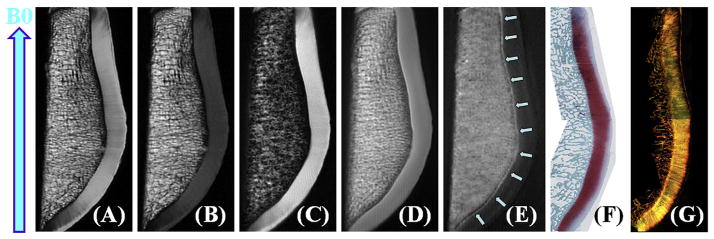
Fig. 1.
High resolution imaging of a cadaveric human patella from the knee joint of a 58-year old male donor with 2D PD-FSE (A), T1-FSE (B), SPGR (C), UTE (D), UTE with echo subtraction (E) and histology (F) as well as PLM (G). Clinical FSE and SPGR sequences show near zero signal for the deep radial and calcified cartilage, which is depicted as a high signal line above the subchondral bone (D and E). Histology and PLM confirmed this patella to be normal with a Mankin score of 0–1 and Vaudey score of 0. B0 field (arrow) is shown for the experimental setup.

