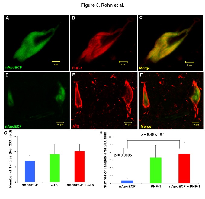
Figure 3. Co-localization of an amino-terminal fragment of apoE within NFTs in hippocampal tissue sections of the Down’s syndrome brain.
(A-C): Representative confocal immunofluorescence double-labeling utilizing the nApoECF antibody (green, Panel A) and PHF-1 (red, Panel B) revealed strong co-localization of the two antibodies within a NFT located in the hippocampus (Panel C). (D-F): Identical to Panels A-C with the exception of AT8 (red) being employed. For Panels A-F, images were captured from the CA1 region of the hippocampus in representative DS-AD cases. (G and H): Quantification of NFTs double-labeled by PHF-1, AT8, and nApoECF. For both panels, data show the number of NFTs labeled with nApoECF alone (blue bar), AT8 (G) or PHF-1 (H) alone (green bars) or those NFTs that were labeled with both antibodies (red bars). NFTs were identified in a 20X field within hippocampal tissue sections by immunofluorescence overlap microscopy (n=3 fields using three different DS cases) ±S.E.M. For Panel G there were no statistical differences between the various test groups. Data indicated that roughly 52.5% of AT8-positive NFTs also contained nApoECF, whereas 53.2% of all identified PHF-1-positive NFTs were labeled with nApoECF.

