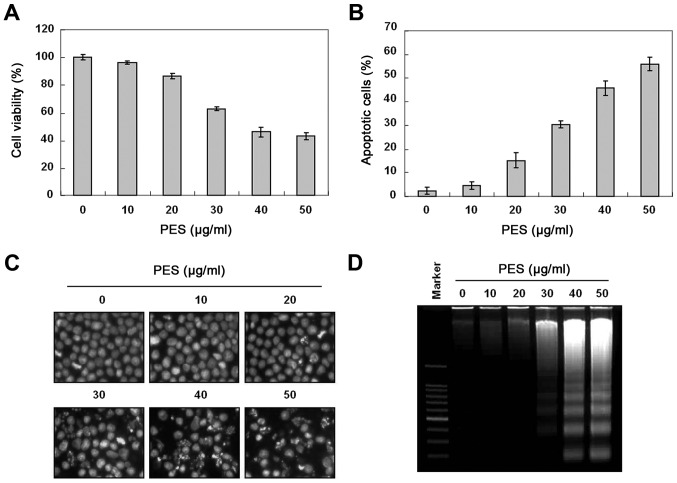
Figure 1.
Growth inhibition by PES in U937 human leukemic cells. U937 cells were incubated at the indicated concentrations of PES for 24 h. (A) The growth inhibition and cytotoxicity of PES were dose-dependent manner. (B) Cell cycle analysis. The cells harboring sub-G1 DNA content represent the fraction undergoing apoptotic DNA degradation by PES treatment. (C) After fixation, the cells were stained with 4′,6-diamidino-2-phenylindole (DAPI) solution to observe apoptotic bodies, which were more frequently observed at higher doses. Stained nuclei were then observed under fluorescence microscopy using a blue filter (magnification, ×400). (D) DNA fragmentation test. A ladder pattern of DNA fragmentation indicates internucleosomal cleavage associated with apoptosis. The data are shown as means ± SD of three independent experiments. *P<0.05 vs. control. PES, polyphenols from Korean E. supina.

