Figure 1.
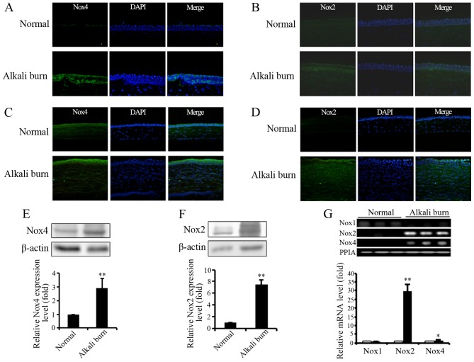
Expression of NADPH oxidase (Nox) isoforms in normal and alkali-burned corneas. (A) Expression of Nox4 was detected by immunofluorescence staining in human normal and alkali-burned corneas. Green indicates the fluorescence signal of Nox4, and blue indicates DAPI fluorescence (original magnification, ×200). (B) Expression of Nox2 in human normal and alkali-burned corneas (original magnification, ×200). (C) Expression of Nox4 and (D) Nox2 was detected by immunofluorescence staining in normal (upper panels) and alkali-burned (lower panels) corneas of mice (original magnification, ×400). The protein level of (E) Nox4 and (F) Nox2 in mouse normal and alkali-burned corneas was examined by western blot analysis. β-actin was used as an endogenous control. The graphs indicate the normalized expression level of Nox4 or Nox2 in corneas. Quantification of Nox4 or Nox2 expression was indicated as the normalization of ratio of Nox4/β-actin or Nox2/β-actin in each sample to control. Data are presented as the means ± SD of at least 3 independent experiments. **P<0.01. (G) RT-qPCR was applied to detect the gene transcription of Nox1, Nox2 and Nox4 in mouse corneas. PPIA was used as an endogenous control. Three independent experiments for each condition were carried out, *P<0.05, **P<0.01 (compared with normal group). The upper panel shows the electrophoresis of the RT-qPCR product of Nox1, Nox2 and Nox4 on an agarose gel.

