Figure 6.
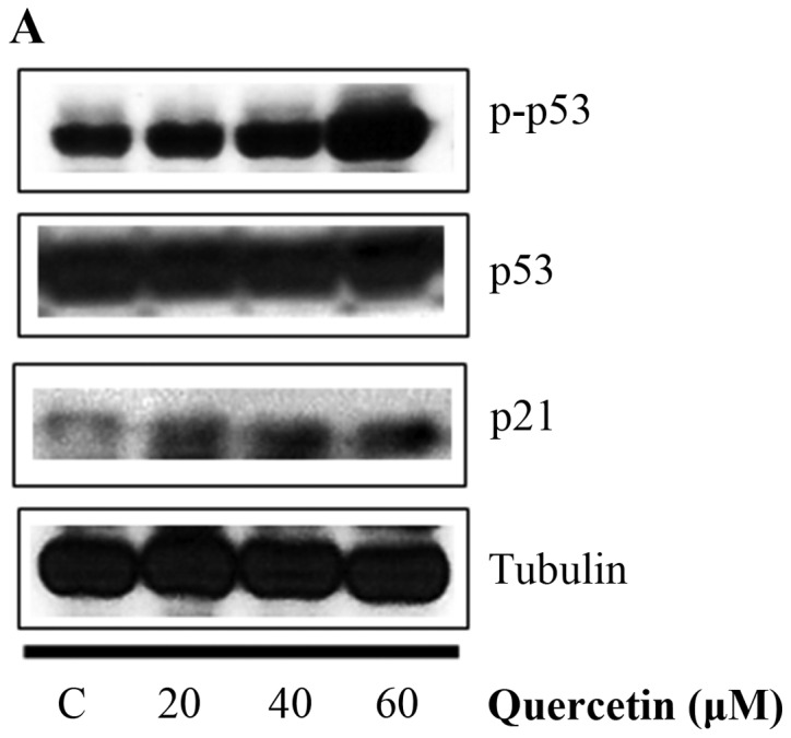
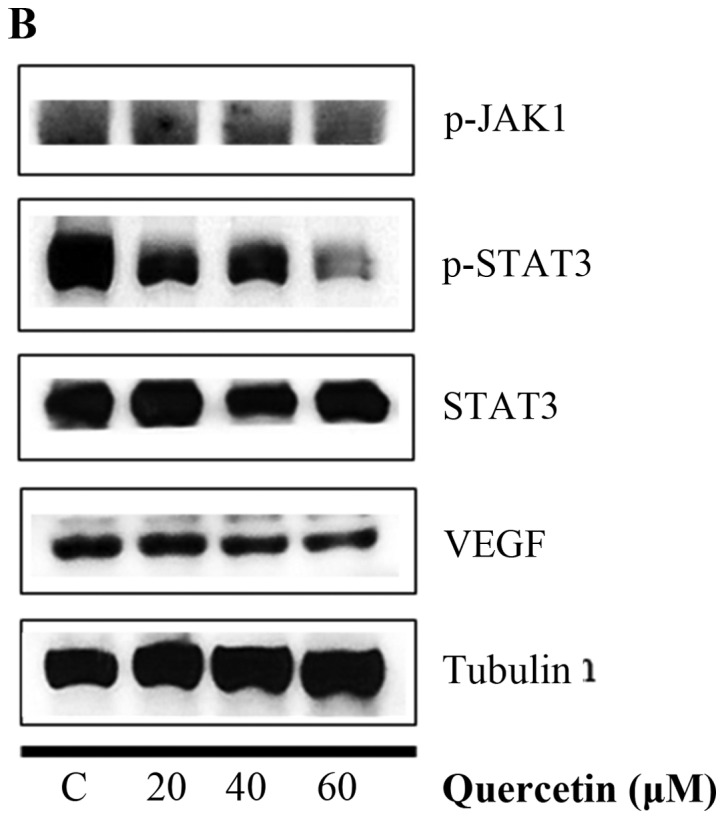
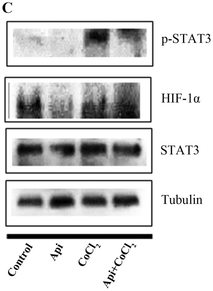
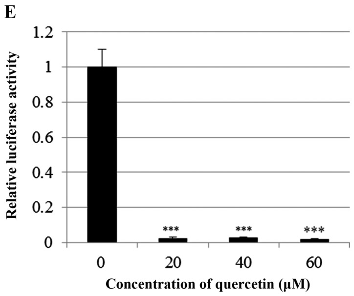
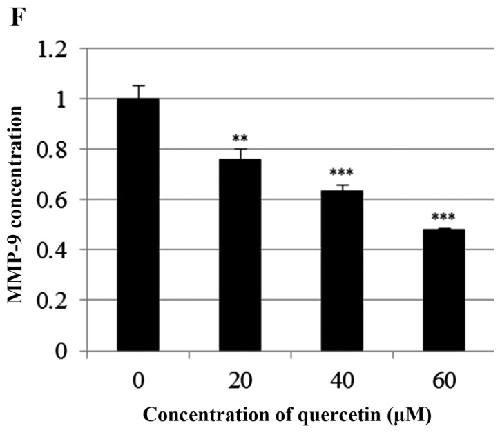
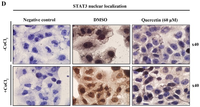
Effect of quercetin on STAT3 activation in BT-474 cells. (A) BT-474 cells were treated with quercetin (0–60 µM) for 24 h. Whole cell lysates were analyzed by western blotting with anti-p-p53, anti-p53, anti-p21 and anti-tubulin antibodies. (B) BT-474 cells were treated with quercetin (0–60 µM) for 24 h. Whole cell lysates were analyzed by western blotting with anti-p-JAK1, anti-p-STAT3, anti-STAT3, anti-VEGF, and anti-tubulin antibodies. (C) BT-474 cells were treated with quercetin (60 µM) for 24 h in the presence or absence of CoCl2 (4 h). Whole cell lysates were analyzed by western blotting with anti-phospho-STAT3, anti-HIF-1α, anti-STAT3, and anti-tubulin antibodies. (D) BT-474 cells were treated with quercetin (60 µM) for 24 h in the presence or absence of CoCl2 and then submitted to immunocytochemistry for detection of nuclear STAT3. The data shown are representative of three independent experiments that gave similar results. (E) BT-474 cells were transiently transfected with p4xM67-TK-luc plasmid containing four copies of the STAT-binding site, treated with quercetin (0–60 µM) and submitted to dual-luciferase assay. (F) BT-474 cells were treated with quercetin (0–60 µM) for 24 h and the intracellular MMP-9 concentration was measured by ELISA. Data are shown as the means of three independent experiments (error bars denote SD). *P<0.05, **P<0.01, ***P<0.001.

