
Figure 1. V2a Brainstem Neurons Project to the Lumbar Spinal Cord and Are Excitatory.
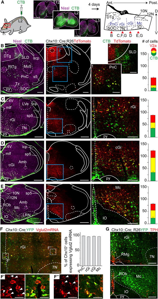
(A) Bilateral injections of the retrograde marker CTB at the 2nd lumbar segment of the spinal cord (left). To the right is a sagittal schematic of the brainstem indicating the approximate rostrocaudal levels shown in the specified panels (red arrows) where CTB-labeled neurons are detected.
(B–E) Transverse hemi-sections at the levels indicated in (A) stained for CTB, Nissl, and V2a neurons (Tdtomato). Right-most pictures are magnifications of the blue boxed area; V2a reticulospinal neurons appear yellow. Bar-graphs show the average number of CTB, V2a, and double-labeled neurons per hemisection (n = 4 animals). Error bars are SEM.
(F) Transverse hemi-section in the rGi indicating Vglut2+ glutamatergic (red) V2a neurons (YFP). Bar-graphs show the average percentage of Vglut2+;V2a neurons (n = 3 animals). Insets in F′ and F″ are magnified views of Vglut2 expression alone (left) and merged with YFP (right) of medially-(F′) and laterally positioned (F″) V2a neurons. White arrowheads indicate co-expression. Error bars are SEM.
(G) Transverse hemi-sections stained for TPH and V2a neurons (YFP). Note the absence of co-expression (n = 4 animals).
Scale bars (in μm): (A): 500, low magnifications in (B)–(E): 500; magnified views in (B)–(G): 200; F′ and F″: 50. Abbreviations used in all figures: 10N: dorsal motor nucleus of vagus; 12N: hypoglossal nucleus; 4V: 4th ventricle; 7N: facial nucleus; Amb: ambiguus nucleus; Gi: gigantocellular reticular nucleus; GiA: gigantocellular reticular nucleus alpha part; icp: inferior cerebellar peduncle; IO: Inferior olive; DTg: laterodorsal tegmental nucleus; LRN: lateral reticular nucleus; LVe: lateral vestibular nucleus; Mc: magnocellular reticular nucleus; mlf: medial longitudinal fasciculus; Mo5: motor trigeminal nucleus; PnC: caudal pontine reticular nucleus; py: pyramidal tract; ROb: raphe obscurus nucleus; RMg: raphe magnus nucleus; RtTg: reticulotegmental nucleus of the pons; s5: sensory root of the trigeminal nerve; scp: superior cerebellar peduncle; SLD: Sublaterodorsal tegmentum; SOC: Superior olivary complex; sp5: spinal trigeminal tract; Tz: nucleus of the trapezoid body. See also Figure S2.