
Figure 7. V2a Stop Neurons Terminate Predominantly in Lamina VII of the Lumbar Spinal Cord.
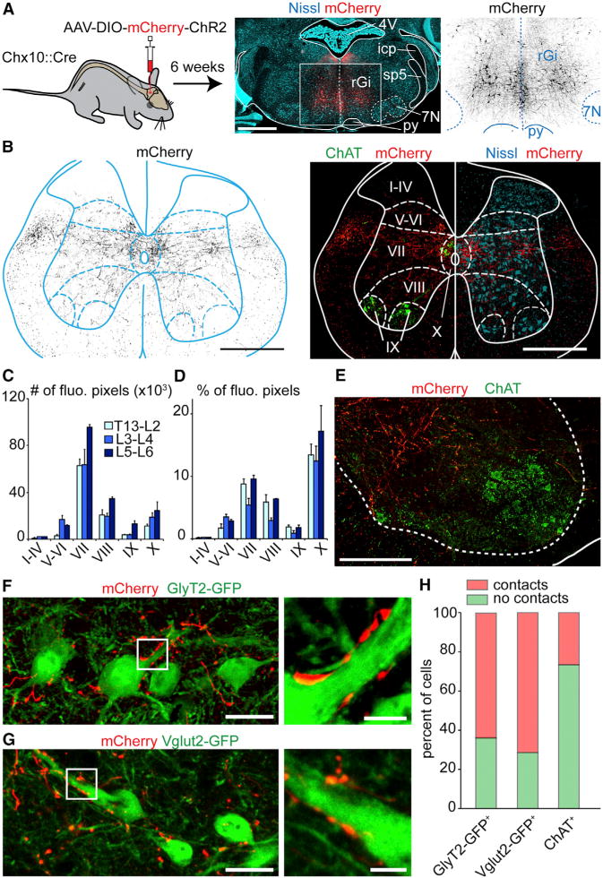
(A) Bilateral injections of a Cre-dependent AAV-hChR2-mCherry-virus (middle: red; right: black) in the rGi of Chx10∷Cre animals.
(B) Transverse L2 spinal cord section of the same animal showing transfected V2a processes (black on the left, red on the right). Rexed’s laminae are delineated using ChAT and Nissl staining.
(C–D) Quantification in one animal of the number (C) or the percent (D) of fluorescent pixels in each lamina at the upper (T13-L1-L2), intermediate (L3–L4), and caudal (L5–L6) lumbar levels.
(E) Magnified view of the descending V2a innervation (red) in the vicinity of ChAT+ motor neurons (green).
(F, G) Similar anterograde labelings on a Chx10∷Cre; GlyT2-GFP (E) or Chx10∷Cre; Vglut2-GFP animal (F) showing putative V2a contacts (red) onto glycinergic and glutamatergic neurons, respectively.
(H) Percent of glycinergic (232 cells), glutamatergic (105 cells), and motor neurons somatas (124 cells) exhibiting no (green) or more than one (red) putative V2a contact.
Scale bars (in μm): (A): 1000, (B): 500, (E): 200, (F, G): 25, insets in (F,G): 5; Error bars in (C) and (D) are SEM; See also Figure S7.