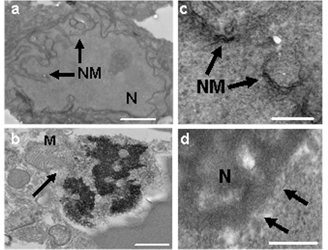
Figure 4.
Ultrastructural analyses of injured cells at 24 h after intracerebral hemorrhage. (a) Normal nucleus (N) and nuclear membrane (NM) of an intact neuron in the contralateral (uninjured) striatum showing intact cellular architecture. (b) Swollen mitochondria (M), nuclear karyorrhexis, and dissolution of the nuclear membrane with mitochondria entry into the nucleus in a cell with necrotic morphology. (c, d) Loss of nuclear membrane integrity consistent with necrosis (arrows indicate nuclear membrane). Scale bars: (a) 2 um, (b) 3 um, (c, d) 300 nm.

