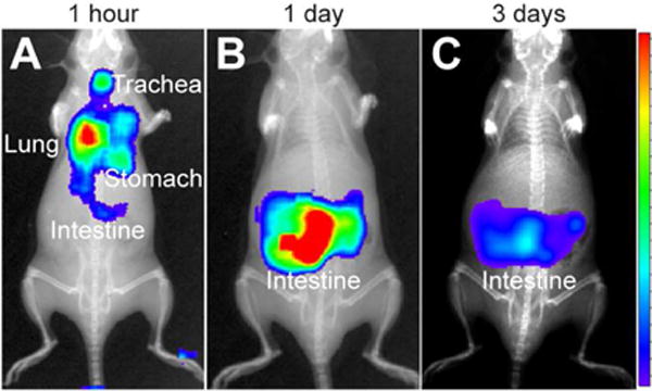
Figure 6.

Ĉerenkov imaging of Dg-cSCK:pDNA nanocomplex clearance. Mice were positioned in the supine position for luminescent imaging of ventral surface after intratracheal administration of 131I-labeled Dg-cSCK:pDNA nanocomplexes (A) immediately post administration, (B) after 3 days and (C) 7 days. The intensity scale is represented on the right.
