Figure 1.
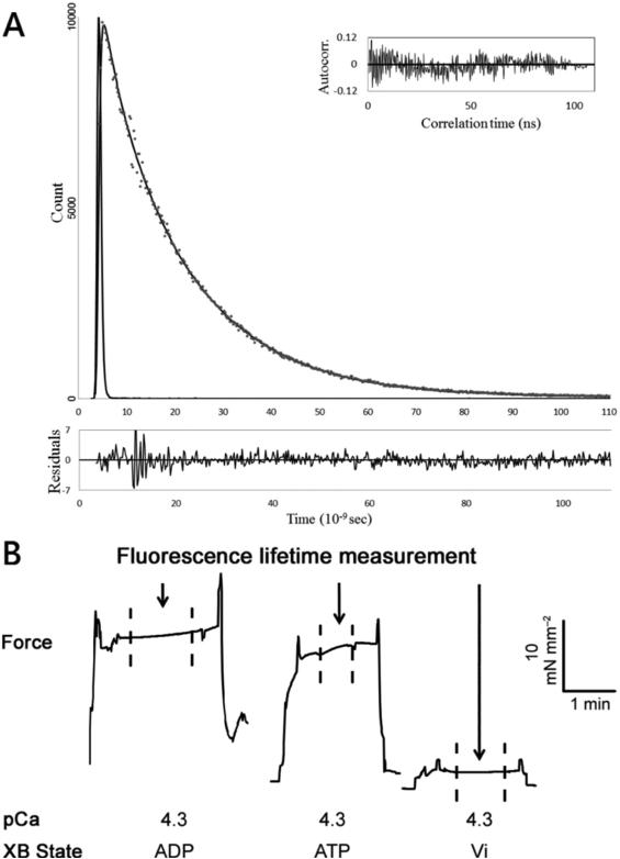
Simultaneous measurements of isometric force and time-resolved fluorescence intensity decay were performed in detergent-skinned cardiac muscle preparations reconstituted with fluorescence donor, cTnI(167C)AEDANS, and nonfluroescent acceptor, cTnC(89C)DDPM. (A) (Upper panel) Representative trace of the In Situ total fluorescence intensity decay of FRET donor in the presence of nonfluorescent acceptor DDPM (grey dots), which was measured at 1.8 μm sarcomere length in the presence of Ca2+ (pCa 4.3). The decay data was fitted with Eq. 1 (black line). The autocorrect (inset) and residual (lower panel) were used to judge the goodness of fit. (B) Fluorescence intensity decays were measured when force had reached steady state as indicated by the arrows and dash lines. Different crossbridge (XB) states are also indicated. The magnitudes of force and time are indicated by the horizontal and vertical bars.

