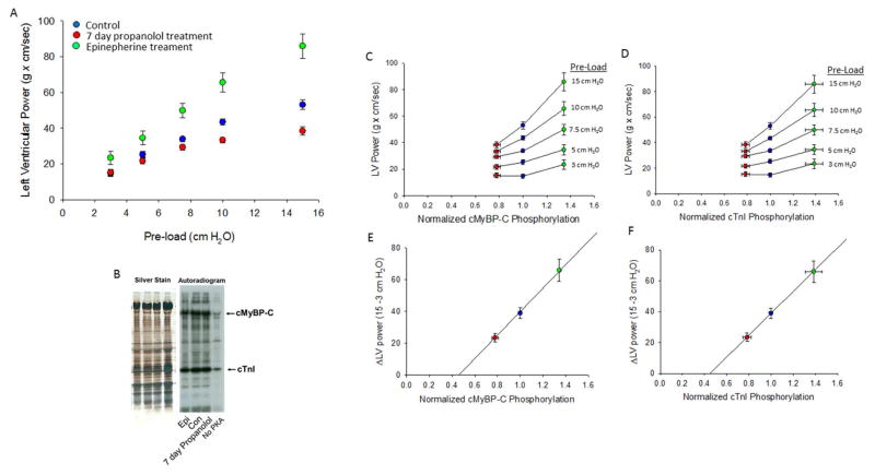
Figure 1. Relationship between isolated rat heart left ventricular (LV) power output and normalized PKA-mediated phosphorylation of cMyBP-C and cTnI.
(A) LV power increased at greater pre-loads. The LV versus pre-load relationship (i.e., ventricular function curve) was most shallow in hearts from rats treated for 7 days with propranolol (n=9), intermediate in hearts from control (untreated) rats (n=14), and steepest in isolated hearts treated with epinephrine (0.1 mM) (n=8). (B). Silver-stained SDS-PAGE and autoradiogram. Cardiac myofibrils were isolated from hearts and treated with (0.1 U/μl) PKA (except last lane) for 45 minutes. PKA-mediated back phosphorylation was assessed by densitometric analysis of exposed bands on film and normalized to protein load from silver stained gel. (C & D) Relationship between LV power and normalized cMyBP-C and cTnI phosphorylation at each pre-load. (E & F) Relationship between change in LV power (ΔLV Power) and normalized cMyBP-C and cTnI phosphorylation.

