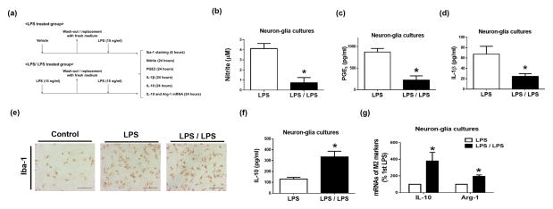
Figure 4. Tolerant microglia display M2-like polarized phenotype.
(a–g) Neuron-glia cultures were pre-treated with vehicle (LPS group) or 15 ng/ml LPS (LPS/LPS group) for 6 hours and then replaced with fresh treatment medium. Additional 6 hours later, LPS (15 ng/ml) was added into the medium again. At 24 hours after LPS re-stimulation, supernatant levels of nitrite (b) was measured with the Griess reagent, pro-inflammatory factors PGE2 (c) and IL-1β (d) and anti-inflammatory cytokine IL-10 (f) were detected by ELISA at different time points indicated in (a). Levels of Iba-1 protein (e) and mRNA of M2 markers IL-10 and Arg-1 (g) were measured by immunocytochemistry (e) and real-time PCR (g), respectively. Asterisk, p<0.05, compared with corresponding LPS-treated cultures without LPS pre-treatment.

