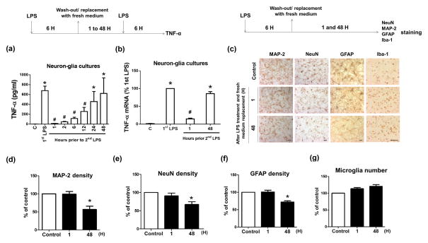
Figure 6. Microglia loss their ability to form ET when surrounding neurons/astroglia were damaged.
(a, b) Neuron-glia cultures were pre-treated with 15 ng/ml LPS for 6 hours. After washed and incubated with fresh media for indicated time, the cultures were treated with LPS and the expression of TNF-α at the level of protein (a) and mRNA (b) was detected 6 hours later. Asterisk, p<0.05, compared with corresponding vehicle-treated control cultures. Number sign, p<0.05, compared with corresponding LPS-treated cultures without LPS pre-treatment. (c–g) Neuron-glia cultures were treated with vehicle or 15 ng/ml LPS for 6 hours and then replaced with fresh medium. Additional 1 or 48 hours later, the expression of MAP-2, NeuN, GFAP and Iba-1 in those cells was measured by immunocytochemistry. (d–f) The density of MAP-2, NeuN and GFAP immunostaining shown in (c) was measured and quantified. (g) The number of Iba-1-immunoreactive microglia of each well was counted under the microscope. Data were shown as the percentage of vehicle-treated control and expressed as the means ± SEM from 5 independent experiments in triplicate. Asterisk, p<0.05 and Number sign, p<0.01, compared with corresponding vehicle-treated control cultures.

