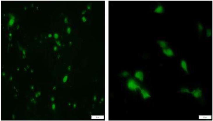
Fig 1.
Immunofluorescent images of the expression of TTSuV1 ORF1 and 2. The swine macrophage cell line 3D4/31 was transfected with TTSuV1 ORF1 (left image) or ORF2 (right image). Protein expression was detected with either a rabbit anti-ORF1 antibody (ORF1) or mouse anti-V5 tag monoclonal antibody (ORF2). Apple green fluorescence in the nucleus for ORF1 and in both the nucleus and cytoplasm for ORF2 is indicative of expression of the relevant protein. Representative images at 24 hrs post-transfection are presented. Fluorescence was not detected in untransfected cells (image not shown).

