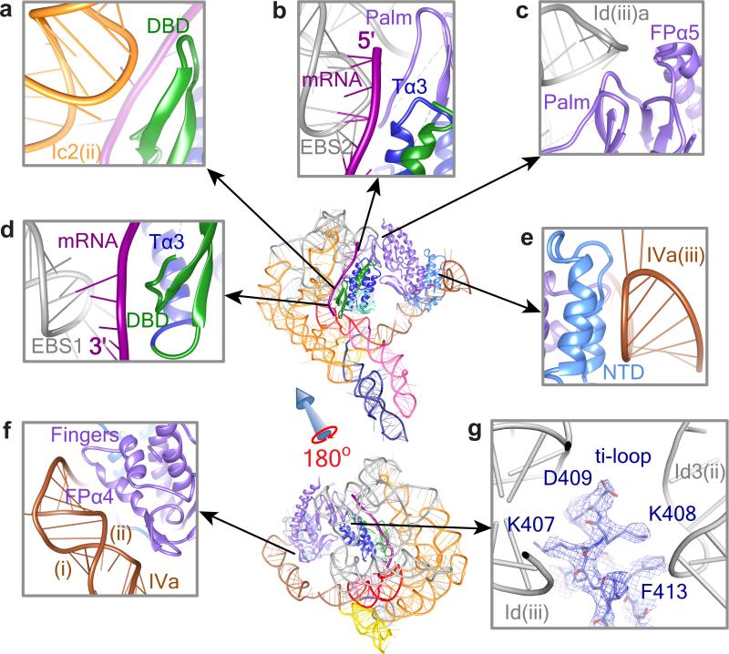
Fig. 4. RNA-Protein Interactions.
Protein-RNA interactions in the RNP complex are shown between Ic2(ii) and DBD (a), between EBS2-IBS2 and Tα3 (b), between Id(iii)a and FPα5 (c), between EBS1-IBS1 and DBD (d), between IVa(iii) and NTD (e), and between IVa(i)&(ii) and FPα4 (f). Panel g shows the ti loop of the thumb domain of LtrA, with well-resolved densities for the bulky side chains of amino acids interacting with apical loops of Id(iii) and Id3(ii). Regions are zoomed in from the thumbnails as indicated by arrows. Abbreviations: T, thumb; FP, fingers-palm. RNA and protein structures are labeled as in Figs. 2b and 3a, respectively.

