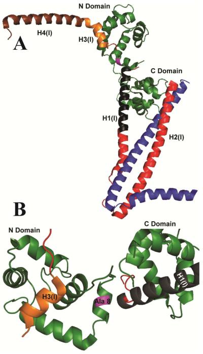
Figure 1. Ribbon diagram of the Ca2+-saturated structure of cardiac Tn showing the site of the mutation (magenta).
A. Peptide backbones are shown for TnC (green), TnI (red, black, gold and brown) and TnT (blue). Residues 150–159 corresponding to the switch region of TnI, H3(I), are shown (gold) at the site of interaction with the hydrophobic patch of TnC. B. The position of alanine 8 (purple) is shown in relation to the H3(I) helix and the lobes of TnC, which form Ca2+-dependent (N-Domain) and Ca2+-independent (C-Domain) contacts with TnI.

