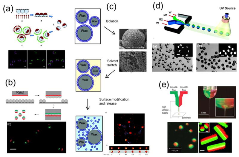
Figure 2.
(a) Top: Schematic illustration showing fabrication of half-coated Janus particles by vapor deposition of gold on silica particles and subsequent surface functionalization. Bottom: Fluorescence images showing proteins conjugated with different fluorescent labels on two hemispheres of the Janus particles. Reprinted with permission from ref. 14 Copyright 2012 American Chemical Society. (b) Top: Schematic illustration of the “sandwich” micro-contact printing method. Bottom: Overlaid epifluorescence images of 3 μm triblock Janus particle that were “printed” with two protein patches of distinctive fluorescence. Reprinted with permission from ref. 67 Copyright 2015 Royal Society of Chemistry. (c) The general process of Janus particles made via the emulsion templating method and scanning electron microscopy (SEM) and fluorescent images of the particles in the process. Reprinted with permission from ref. 68 Copyright 2006 American Chemical Society. (d) Top: Schematic illustration of the microfluidic fabrication of Janus particles. Bottom: Brightfield and fluorescence images of the Janus particles. Reprinted with permission from ref. 71 Copyright 2006 American Chemical Society. (e) Top: Experimental setup of the electrohydrodynamic co-jetting technique for synthesizing biphasic Janus particles. Bottom: Overlaid fluorescence images of the generated Janus spheres and cylinders. Reprinted with permission from ref. 77 Copyright 2005 Nature Publishing Group and ref. 78 Copyright 2009 Wiley-VCH Verlag GmbH & Co. KGaA.

