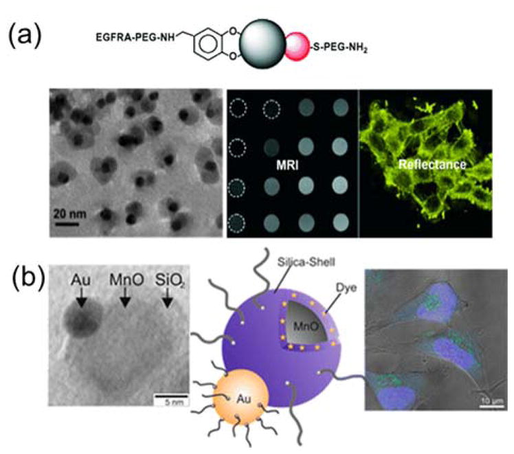
Figure 4.

Janus nanoparticles for dual bioimaging in cells. (a) Top: Schematic illustration of surface functionalization of the Au-Fe2O3 Janus nanoparticles. Bottom: TEM and MRI images of the nanoparticles, and reflectance image showing cells labeled with the nanoparticles. Reprinted with permission from ref. 134 Copyright 2008 Wiley-VCH Verlag GmbH & Co. KGaA. (b) From left to right: TEM image showing an Au-MnO Janus nanoparticle, schematic illustration of the functionalization of the Janus nanoparticle, and confocal laser scanning microscopy image of Hela cells stained with the fluorescently labeled Au-MnO Janus nanoparticles. Reprinted with permission from ref. 139 Copyright 2014 American Chemical Society.
