Figure 1.

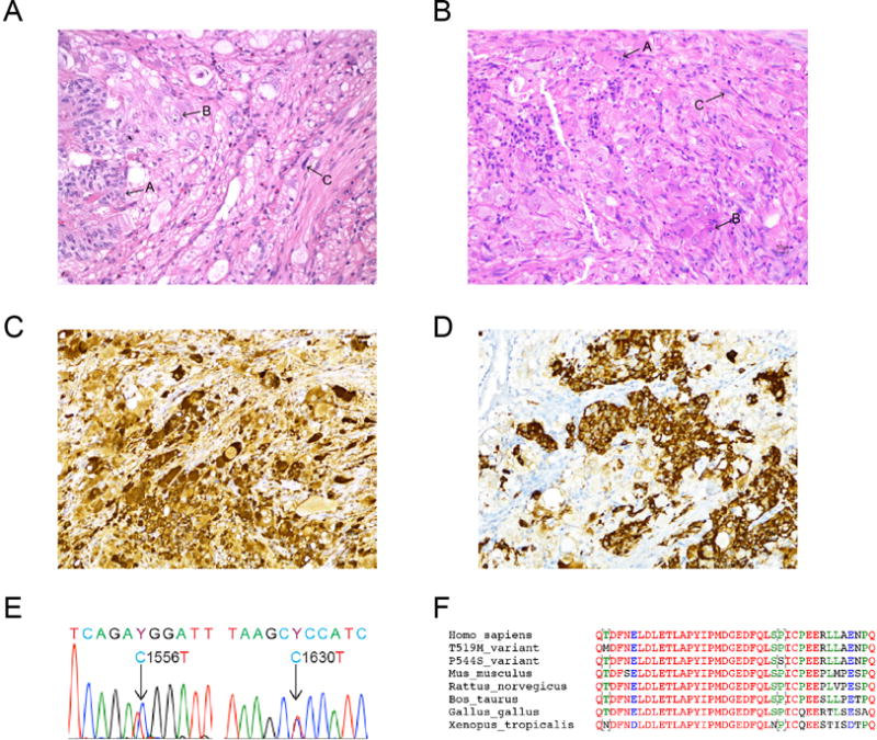
A–B. Histopathological features of GPGLs from patient 1 (A) and patient 2 (B). In both tumors, the neoplastic cells were arranged in solid nests, trabeculae and fascicules with areas of epithelioid (arrow A), ganglion-like (arrow B), and spindle (arrow C) cells. Hematoxylin & eosin stain, magnification × 20. Positive immunohistochemical stains for chromogranin A (C) and somatostatin (D) in the tumor from patient 1, magnification × 20. Both GPGLs were negative for tyrosine hydroxylase (ImmunoStar) immunohistochemistry (data not shown). E. Genomic DNA sequencing of HIF2A exon 12 nucleotides 1551–1561. Heterozygous T519M (patient 1) and P544S (patient 2) somatic mutations were identified in both GPGLS. No HIF2A mutations were found in the blood or gallbladder tissue of patient 1. F. Alignment of amino acid sequence of HIF-2α residues 1625–1635 in humans, mice, rats, ox, chickens, and frogs.
