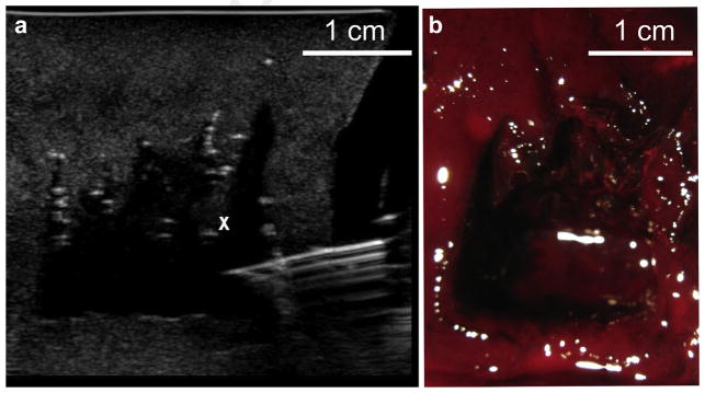
Fig. 4.
A large boiling histotripsy liquefied lesion was produced in a hematoma sample by translating the focus of the 1.5 MHz transducer in a 2 × 2 cm grid in 2-mm steps. (a) B-mode image of the fine-needle (18-gauge) aspiration of lysate from the cavity. The cavity appears as a hypoechoic region; note the irregular filaments protruding from the side of the void (the white “x” denotes one of them). These filaments are due to the incomplete merging of the distal parts of the individual liquid lesions. (b) Photograph of the bisected treated hematoma sample.

