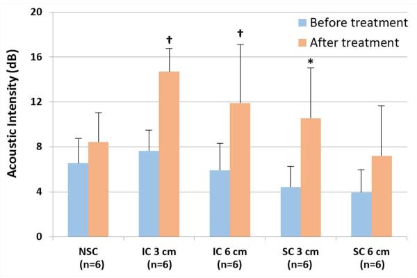
Figure 3. Demonstration of the increase in skeletal MBV with inertial cavitation (IC) inducing ultrasound treatments.
Error bars depict standard deviation. SC = stable cavitation; NSC = nondestructive stable cavitation; 3 cm = 3 cm phantom; 6 cm = 6 cm phantom. *<0.05 compared to before treatment; †<0.01 compared to before treatment. n = 6 for each group.

