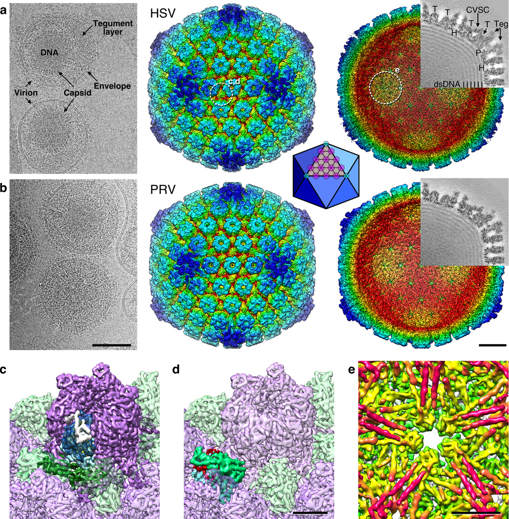
Figure 2. HSV-1 and PRV density maps.
(a) HSV-1 and (b) PRV. Left, representative portions of cryo-micrographs showing intact virions from which capsid images were collected. Elements of the virion are marked in (a). Scale bar 1000 Å. Surface renditions of the capsid reconstructions colored by radius are shown viewed from the exterior (center) and interior (right) after computational removal of internal density. White dashed circles mark the locations highlighted in panels c–e, as indicated. Superimposed on each interior view is a grey-coded section (protein is dark) corresponding to the sectioning plane and with structures indicated as follows: “H”, hexon; “P”, penton; “T”, triplex. Density attributed to tegument is indicated as “Teg”, and to the CVSC molecule is arrowed. Scale bar 200 Å. Inset: a diagram of the icosahedral lattice geometry with triangulation number T=16 indicating the locations of hexons (purple) and pentons (light blue). (c) Close-up view of a hexon (purple) with the domains of one VP5 subunit colored as dark blue (upper domain), light blue (middle domain) and dark green (lower domain). The VP26 subunit bound to the VP5 tip is colored white. (d) Close-up view of a triplex molecule with proposed subunit segmentation of magenta, green and blue. (e) Close-up view beneath the HSV-1 penton, revealing striking pairs of tubes (magenta). Scale bars 50 Å.

