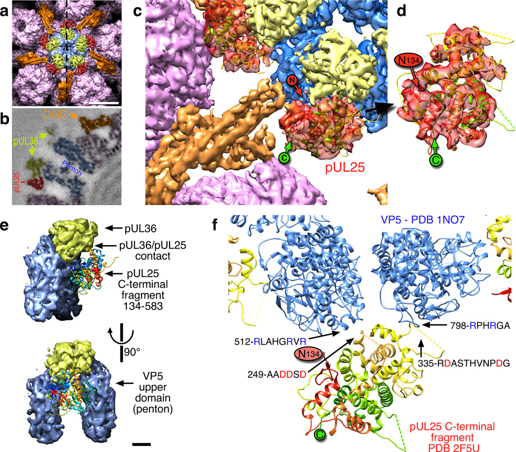
Figure 7. pUL25 – localization and contacts.
(a) Surface view above the penton (blue) including hexons (purple), CVSC pUL17 and the N-terminal domain of pUL25 (orange), and the CVSC C-terminal domain of pUL25 (red). The dashed line indicates the sectioning plane in (b) which is colored similarly. Scale bar 100 Å. (c) Atomic model of the HSV-1 pUL25 C-terminal fragment (residues 134–580; PDB 2F5U8) fit into the PRV density map in the peri-pentonal region of the CVSC molecule (red). The location of the N-terminal residue 134 (“N” in red circle) is adjacent to the remainder of the CVSC density (orange). (d) An enlargement of the fit in an orientation where the correspondence is more readily apparent. Dashed lines indicate mobile loops not resolved in the crystal structure. (e) Contacts between pUL25 C-terminal domain (ribbon diagram) and two VP5 subunits of the penton (blue) as well as the pUL36 density above the penton (yellow). Scale bar 20 Å. (f) Pseudo-atomic model of the pUL25–VP5 interface highlighting the complimentary charges of the contacting loops.

