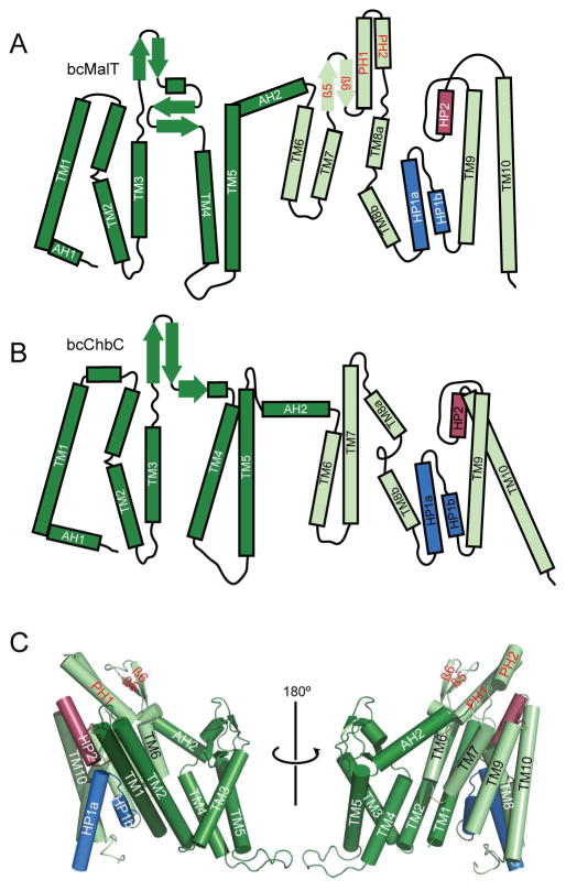
Figure 2. Structure of bcMalT.
a–b. Topology diagrams of bcMalT (a) and bcChbC (b) oriented with the periplasmic side on top. The N-terminal dimerization domain and C-terminal transport domain are colored dark and light green, respectively; HP1 and HP2 are colored marine and raspberry. Structural elements in the transport domain not shared with bcChbC are indicated with red labels, and include a periplasmic loop following TM7 containing a two-stranded antiparallel β-sheet (β5 and β6) and two additional α-helices (periplasmic helices PH1 and PH2). c. Protomer of bcMalT in two orientations, colored according to the scheme in panel a. Hairpin loops HP1 and HP2 are shown in marine and raspberry.

