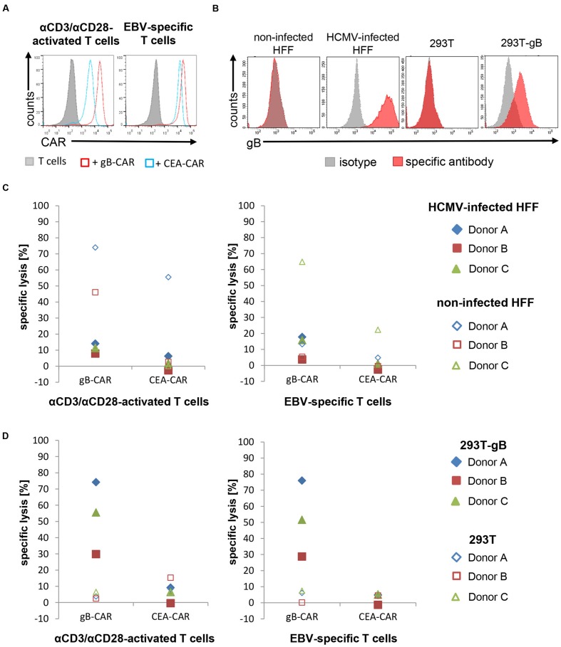
FIGURE 1.
Lysis of HCMV-infected HFF by CAR-T cells is strongly reduced. (A) The histograms shows the expression of the different CARs in αCD3/αCD28-activated or EBV-specific T cells, respectively, 20 h after mRNA electroporation of a representative donor (EBV-specific T cells n = 6; 4 donors; αCD3/αCD28-activated n = 3; 3 donors). (B) Flow cytometric analysis of the gB-expression in 293T cells, gB-expressing 293T (293T-gB) cells, non-infected and HCMV-infected HFF. Shown is a representative histogram of one experiment (n = 3). (C,D) The diagrams show the lytic potential of redirected αCD3/αCD28-activated or EBV-specific T cells targeting HCMV-infected HFF (C) or gB-expressing 293T (293T-gB; (D) analyzed in a 4 h Eu release assay (E:T-ratio referring to CD8pos T cells = 25:1). 293T cells, non-infected HFF were used as target cell controls, CEA-CAR-expressing EBV-specific T cells were used as controls for CAR-T cells. (B–D) HCMV-infected HFF in all shown experiments were used at 4 days after infection (AD169, MOI 5).

